Di attach dong imagenya,
Gag tampil nih.
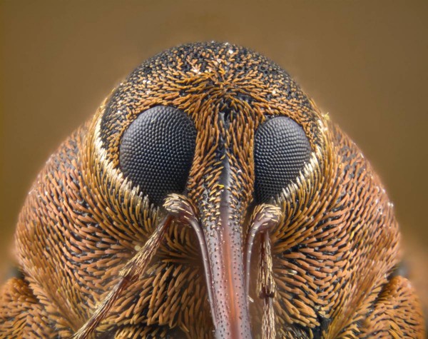
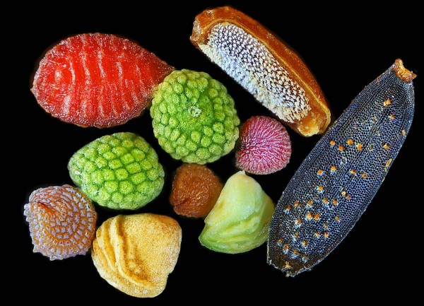
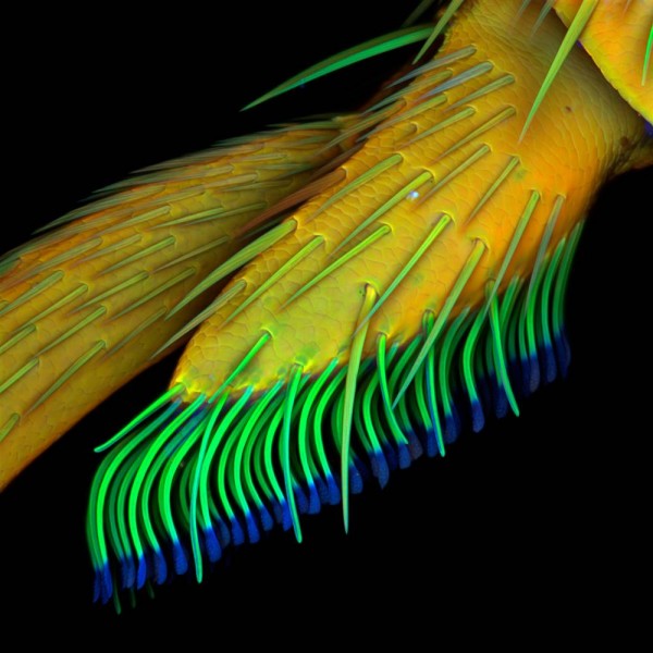
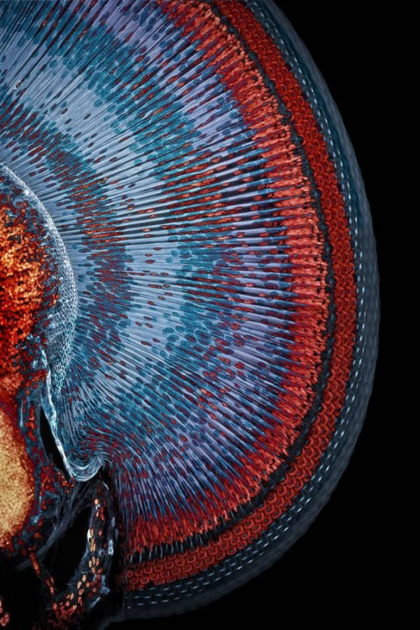
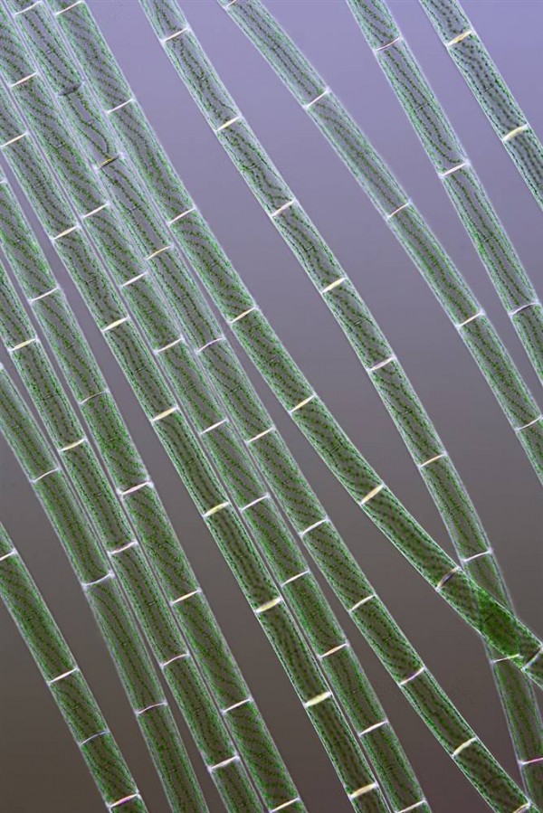
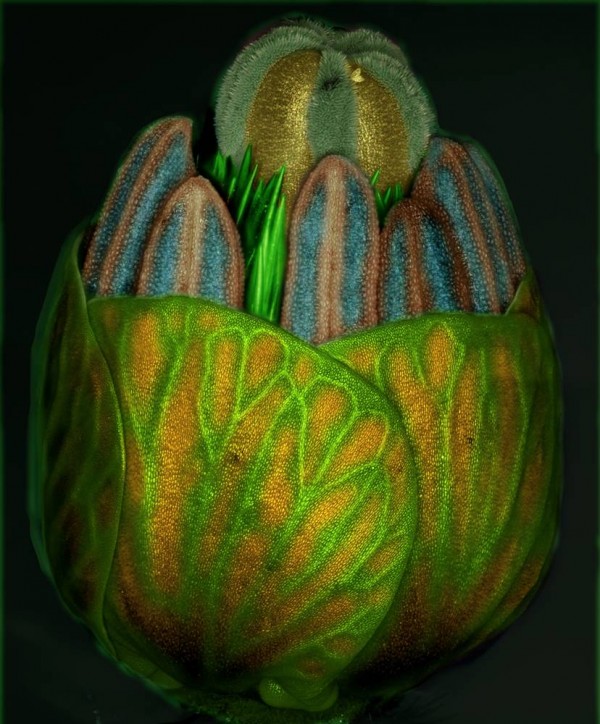
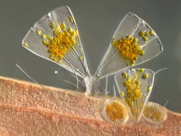
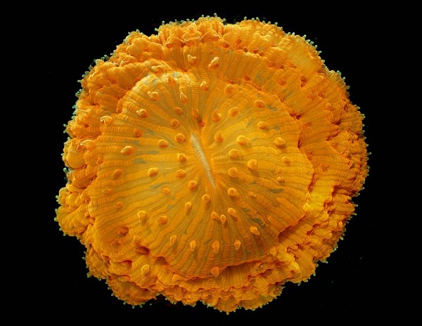
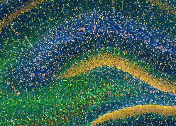
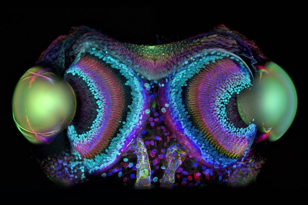
NB : We will not accept any issue about quality, so please send out your best
quality.
Terima Kasih dan Hormat Kami,
Nur Ahmad Faizi (Izzy)
Purchasing - Development
[cid:[email protected]]PT. SHYANG YAO FUNG Plant B
Address : Jl. Industri Raya Blok D No.2
Tel         : 021-59300888 Ext 7706 / Ph: 0857-1023-8591/021-91164937
Fax        : 021-59301777
Email     : [email protected]<mailto:[email protected]>
===========================================================
"Narrated Abu Huraira(r.a): The Prophet said:"... whoever fasts during Ramadan
out of sincere faith and hoping to attain Allah's rewards, then all his past
sins will be forgiven." (Sahih Al-Bukhari)."
P   Think of our planet. Please consider the environment before printing this
email


________________________________
From: [email protected] [mailto:[email protected]] On
Behalf Of aga madjid
Sent: Tuesday, August 02, 2011 8:04 AM
Subject: ~ aga ~ Extraordinary Microscope Images


10. Weevil Head
This is the 10th-place picture, by British photographer Laurie Knight, shows
the face of a weevil (possibly Curculio nucum or Curculio glandium). The image
was captured using a lighting technique known as episcopic illumination.
[http://thewondrous.com/wp-content/uploads/2010/11/Weevil-Head-600x475.jpg]
The Olympus BioScapes International Digital Imaging Competition honors the
world's most extraordinary microscope images of life science subjects.
9. Wildflower seeds
China's Yanping Wang won ninth place in the 2010 Olympus BioScapes competition
for this image of wildflower seeds, captured using brightfield reflected light.
[http://thewondrous.com/wp-content/uploads/2010/11/Wildflower-seeds-600x433.jpg]
8. Beetle Leg
German researcher Jan Michels' eighth-place image shows a lateral view of the
adhesive pad on the leg of a beetle (Clytus sp.). The view was captured using
autofluorescence.
[http://thewondrous.com/wp-content/uploads/2010/11/Beetle-leg-600x600.jpg]
7. Damselfly's Eye
Germany's Igor Siwanowicz won seventh place in the Olympus BioScapes
competition for this view of the eye of a damselfly. This projection of a
series of confocal microscope images shows the regular, crystal-style
architecture of the eye of Enallagma cyathigerum. The area covered in the photo
measures about 0.6 by 0.8 millimeters, or 0.02 by 0.03 inches.
[http://thewondrous.com/wp-content/uploads/2010/11/Damselflys-eye-600x899.jpg]
6. Spirogyra
Polish biotech researcher Jerzy Gubernator took this extreme close-up of
Spirogyra algae using brightfield and polarized light. The photomicrograph won
the sixth-place prize in the 2010 Olympus BioScapes competition.
[http://thewondrous.com/wp-content/uploads/2010/11/Spirogyra-600x899.jpg]
5. Primordium
This picture by Iranian horticulturist M. Reza Dadpour shows the primordium
(bud) of the weedy flower Tribulus sp. during its final stages of development.
More than 100 images, focused at different depths, were composed to produce
this view. The image won fifth prize in the 2010 Olympus BioScapes competition.
[http://thewondrous.com/wp-content/uploads/2010/11/Primordium-600x724.jpg]
4. Diatoms
Fanlike diatoms, representing the species Licmophora juegensii, have latched
onto red algae in this fourth-place picture by Germany's Wolfgang Bettighofer.
Licmophora cells are able to move and, supported through light sensors, locate
a place with suitable light exposure. Then they produce a sticky stalk that
keeps them attached to their home base. The sample was collected from brackish
water in the Baltic Sea.
[http://thewondrous.com/wp-content/uploads/2010/11/Diatoms-600x451.jpg]
3. Coral
The third-place image in the Olympus BioScapes competition shows a solitary
coral (Fungia sp.). The tentacle tips, called acrospheres, are visibly
enhanced. This picture was taken by James Nicholson, a coral researcher in
South Carolina.
[http://thewondrous.com/wp-content/uploads/2010/11/Coral-600x464.jpg]
2. Rat Brain
California's Thomas Deerinck won second place in the Olympus BioScapes
competition for this image of a structure known as the hippocampus, within the
brain of a rat. The tissue was stained to reveal the distribution of glial
cells (blue), neurofilaments (green) and cell nuclei (yellow).
[http://thewondrous.com/wp-content/uploads/2010/11/Rat-brain-600x431.jpg]
1. Daddy Longlegs
Germany's Igor Siwanowicz won first place in the Olympus BioScapes competition
for this image, showing the eyes of a spiderlike bug known as a Daddy Longlegs
or Harvestman. The picture has been color-coded to reflect depth, and shows the
lenses (two large ovals), the retinas and the optic nerves.
[http://thewondrous.com/wp-content/uploads/2010/11/Daddy-longlegs-600x400.jpg]


--
".... I am the KING to my own UNIVERSE that Rule my MIND, BODY and SOUL !!!
...."

- Aga Madjid -

--
you have this email because you join to "aga-madjid" GoogleGroups.
to post emails, just send to :
[email protected]
to join this group, send blank email to :
[email protected]
to quit from this group, just send email to :
[email protected]
please visit to www.facebook.com/aga.madjid,
add my Yahoo Messenger at [email protected] or
add my twitter @aga_madjid
thanks for joinning this group.
<<inline: image001.jpg>>